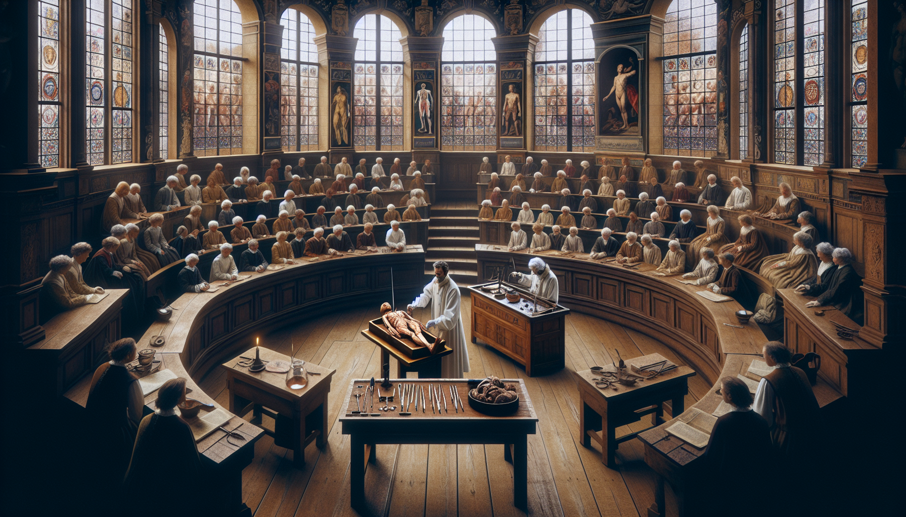
In the dimly lit chambers of 17th-century Europe, where the mysteries of the human body were as enigmatic as the distant stars, a curious fusion of art and science began to unfold. As the Renaissance spirit continued to ripple through the corridors of knowledge, a fascination with the human form took center stage. This was a time when public dissections emerged as both a spectacle and a profound educational endeavor, capturing the imagination of artists, scientists, and the general populace alike. The intricate illustrations born from these dissections have survived through centuries, offering us a glimpse into the scientific curiosity and cultural dynamics of an era long past.
Public dissections were not merely medical exercises; they were theatrical events that drew crowds from all walks of life. In an age when the line between science and spectacle was intriguingly blurred, these dissections provided rare opportunities for ordinary citizens to peek inside the human body, which was considered a sacred vessel. Artists, commissioned to capture these events, produced illustrations that are as much works of art as they are scientific documents. These images, brimming with meticulous detail and artistry, reveal the layers of fascination and trepidation that accompanied the exploration of human anatomy. But what drove this public interest in dissections? How did the artists of the time contribute to the dissemination of anatomical knowledge through their work?
In this deep dive into the past, we will uncover the multifaceted roles that these illustrations played in the 17th century. We will explore how artists like Andreas Vesalius and his successors not only documented the dissections but also influenced the way people perceived the human body and its functions. These illustrations were more than mere depictions; they were educational tools, crafted to convey complex scientific concepts to both learned scholars and curious onlookers. As we journey through the pages of history, we’ll examine the intricate balance between scientific accuracy and artistic expression that these works encapsulate, and how they continue to inspire modern interpretations of the human form.
The impact of these illustrations extended beyond the scientific community. They were instrumental in shaping public opinion and understanding of anatomy and medicine. This article will delve into how these images helped demystify the human body, challenging long-held superstitions and paving the way for a more empirical approach to medicine. We will also consider the broader cultural implications of these dissections and their illustrations, reflecting on what they tell us about the values and beliefs of 17th-century society. Were these public dissections an early form of democratizing knowledge, or did they reinforce existing social hierarchies?
Join us as we unravel the stories behind these captivating illustrations, exploring their significance in both the art and science worlds. Through this exploration, we’ll gain insight into how these historical practices laid the groundwork for modern anatomy and art, and how they reflect the enduring human quest to understand the body and its mysteries. This journey through the past promises to be as enlightening as it is intriguing, shedding light on the intricate dance between observation, representation, and interpretation that continues to shape our understanding of the human condition. 🧠📜
The Fascinating World of 17th Century Public Dissections
The 17th century marked a pivotal era in the history of medicine, characterized by a burgeoning interest in anatomy and the human body. Public dissections, often held in amphitheater-like settings, became both a scientific endeavor and a form of public spectacle. Illustrations from this time provide a vivid depiction of these events, offering insights into the societal and cultural milieu of the period. The detailed and often dramatic representations serve not only as historical records but also as artistic achievements that continue to intrigue scholars and enthusiasts alike.
Public dissections were part of a broader movement toward empirical investigation, a hallmark of the scientific revolution. These dissections were usually performed by leading anatomists of the time and were attended by a wide array of spectators, including medical students, artists, and the general public. The illustrations produced during these dissections were instrumental in disseminating anatomical knowledge across Europe, influencing both the medical community and the public’s understanding of the human body.
One of the most famous anatomical theaters was in Leiden, Netherlands, which hosted numerous dissections attended by large audiences. The events were meticulously documented, with illustrations capturing the intensity and complexity of the dissections. These images serve as a testament to the evolving scientific methods of the time and highlight the intersection of art and science in disseminating knowledge. Furthermore, they provide a window into the cultural attitudes toward death and the human body during the 17th century.
Historical Context of 17th Century Public Dissections
The 17th century was a period of great change and exploration in many fields, including medicine. This era saw the gradual shift from reliance on ancient texts to a more empirical approach to understanding the human body. Anatomical theaters became the epicenter of this transformation, where public dissections played a crucial role in advancing medical knowledge. These theaters were often affiliated with universities and were designed to accommodate a large number of spectators, reflecting the public’s growing interest in science and anatomy.
The illustrations from these dissections offer a unique perspective on the period’s medical practices and societal norms. They reveal not only the anatomical details that were of interest to physicians but also the theatrical nature of the dissections themselves. These events were often elaborate affairs, complete with invitations, announcements, and even refreshments for attendees. The illustrations capture the atmosphere of these dissections, portraying the blend of education and entertainment that defined them.
The cultural significance of these illustrations cannot be overstated. They reflect a time when the boundaries between science and spectacle were blurred, and when the exploration of the human body was both a source of fascination and a subject of artistic expression. These images continue to captivate modern audiences, offering a glimpse into the mindset and values of the 17th century.
The Role of Illustrations in Disseminating Anatomical Knowledge
Illustrations from 17th-century public dissections were vital in spreading anatomical knowledge. These images were not merely artistic interpretations but were often created with a high degree of accuracy and attention to detail. Artists would collaborate closely with anatomists to ensure the illustrations were scientifically valid, which made them valuable resources for medical education. The proliferation of these images helped standardize anatomical knowledge across Europe and facilitated the exchange of ideas among scholars.
These illustrations served several purposes. Primarily, they were educational tools used to supplement live dissections and lectures. For students and physicians who could not attend dissections in person, the illustrations provided an alternative means of learning. Additionally, the images were used in published anatomical atlases, which became essential references for medical practitioners. These atlases were often richly illustrated, combining scientific precision with artistic flair, and were highly sought after by medical professionals and laypeople alike.
In addition to their educational value, the illustrations also played a role in shaping public perceptions of medicine and science. By depicting dissections in a way that was accessible and engaging, these images helped demystify the human body and the process of dissection. They contributed to a broader understanding and acceptance of anatomical studies, which in turn supported the advancement of medical science.
Artistic Techniques and Styles in Anatomical Illustrations
The artistic techniques used in 17th-century anatomical illustrations varied widely, reflecting the diverse backgrounds and skills of the artists involved. Many illustrations were highly detailed and precise, utilizing techniques such as engraving and etching to create clear and accurate representations of anatomical structures. These methods allowed for the production of high-quality images that could be reproduced and distributed widely.
Some artists approached anatomical illustration with a more artistic or expressive style, incorporating elements of drama and emotion into their work. These images often depicted dissections as grand spectacles, with dramatic lighting and composition emphasizing the theatrical nature of the events. This approach not only captured the scientific details but also conveyed the cultural and emotional impact of the dissections.
Furthermore, the interplay between text and image was crucial in these illustrations. Descriptive annotations often accompanied the images, providing context and explanation for the anatomical details depicted. This combination of visual and textual information made the illustrations an effective educational tool, bridging the gap between art and science.
Comparative Analysis of Prominent 17th Century Anatomical Illustrations
When examining 17th-century anatomical illustrations, it is evident that there was a wide range of styles and approaches. Some illustrations focused on the technical and educational aspects, while others emphasized the artistic and dramatic elements. A comparative analysis of these images reveals both the diversity and the commonalities that defined anatomical illustration during this period.
| Illustration Aspect | Technical Focus | Artistic Emphasis |
|---|---|---|
| Detail | Highly precise, emphasizing anatomical accuracy | Less focus on precision, more on emotional impact |
| Composition | Structured and methodical layout | Dramatic and dynamic arrangement |
| Purpose | Educational, primarily for medical students | Entertainment, appealing to a broader audience |
As you can see in the table above, the illustrations with a technical focus were characterized by their precision and methodical composition. These images were primarily intended for educational purposes, serving as valuable resources for medical students and professionals. They often included detailed annotations and were used in conjunction with lectures and dissections to enhance learning.
On the other hand, illustrations with an artistic emphasis prioritized drama and emotional impact. These images were designed to capture the public’s imagination, drawing viewers into the spectacle of the dissection. While they may not have been as precise in terms of anatomical detail, their artistic qualities made them memorable and engaging, appealing to a wider audience beyond the medical community.
Case Studies of Notable Illustrations
To further understand the impact and significance of 17th-century anatomical illustrations, it is useful to examine specific examples in detail. One such case is the work of Andreas Vesalius, whose illustrations set a new standard for anatomical accuracy and detail. His landmark publication, “De humani corporis fabrica,” included detailed engravings that were both scientifically rigorous and artistically sophisticated, establishing a new benchmark for anatomical illustration.
Another notable example is the work of Rembrandt, who, although not an anatomist, captured the drama and intensity of public dissections in his famous painting “The Anatomy Lesson of Dr. Nicolaes Tulp.” This painting, while not an anatomical illustration per se, exemplifies the intersection of art and science, depicting a dissection as a theatrical event. It highlights the role of public dissections as both educational and social occasions, and its dramatic composition and lighting underscore the spectacle of the scene.
For a visual exploration of these fascinating illustrations, you can watch the video “Exploring Historical Anatomy Art” by Artful Science on YouTube: Link to Video. This video delves into the artistic and scientific aspects of anatomical illustrations, providing further insight into their historical context and significance.
Conclusion
Unveiling the Past: Intriguing 17th Century Illustrations of Public Dissections has taken us on an enlightening journey through a fascinating era where art and science intersected in profound ways. This exploration not only sheds light on the artistic and scientific endeavors of the 17th century but also provides a unique perspective on how these public dissections were perceived culturally and socially. By delving into the intricacies of these illustrations, we gain a deeper understanding of the historical context and the advances in anatomical knowledge that shaped modern medicine.
Throughout the article, we have explored the significance of these illustrations as a historical record. The detailed engravings and drawings from this period are not merely artistic expressions but are also vital documents that reflect the burgeoning curiosity and the thirst for knowledge that characterized the Renaissance and the Enlightenment periods. They serve as a testament to the evolving understanding of human anatomy and the shift towards empirical observation and scientific inquiry. The illustrators of the time, often collaborating with anatomists, played a crucial role in disseminating this newfound knowledge, bridging the gap between science and the public.
Moreover, the cultural and social dimensions of these public dissections and their illustrations have been underscored. Public dissections were events that drew large crowds, serving as both educational opportunities and public spectacles. The illustrations capture the dual nature of these events, highlighting the tension between the educational aspirations and the macabre curiosity of the audience. These gatherings were a reflection of societal attitudes towards the body, death, and the quest for knowledge, and the illustrations serve as a window into these complex dynamics.
In examining the artistic techniques employed in these illustrations, we have seen how artists of the 17th century utilized various methods to depict anatomical details with remarkable accuracy. This required a profound understanding of both artistic principles and anatomical science, showcasing the interdisciplinary nature of the work. These illustrations were not only tools for education but also works of art that conveyed the beauty and complexity of the human form. The skill and precision involved in creating these images underscore the respect and reverence held for the human body, even in death.
The technological and methodological advances of the time, as depicted through these illustrations, laid the groundwork for future generations. The integration of art and science in the study of anatomy during this period was a precursor to modern medical illustration, which continues to play a crucial role in medical education today. By examining these historical illustrations, we appreciate the foundation upon which contemporary medical practices are built and recognize the enduring legacy of these early contributions to science and art.
As we conclude our exploration, it is essential to reflect on the lasting impact of these 17th-century illustrations of public dissections. They are more than historical artifacts; they are a reminder of humanity’s relentless pursuit of knowledge and understanding. They challenge us to consider how far we have come in our scientific endeavors and inspire us to continue exploring the unknown. In a world where technology rapidly advances, the lessons from the past remind us of the importance of curiosity, interdisciplinary collaboration, and the human capacity for innovation.
We invite you, dear reader, to further engage with this topic by exploring additional resources and sharing your thoughts and insights. By doing so, you contribute to the ongoing dialogue about the intersection of art, science, and history. Consider how these historical perspectives can inform and enrich our understanding of contemporary issues in medicine and art.
If you found this article intriguing, please share it with others who might be interested in uncovering the fascinating history of anatomical illustration. We encourage you to reflect on how the themes discussed can be applied in various fields today, from education and research to art and cultural studies. Your engagement and curiosity are vital in keeping the spirit of inquiry alive.
For those interested in delving deeper into this topic, we recommend exploring additional academic articles and historical archives. Websites such as the Wellcome Collection (https://wellcomecollection.org/) and the Digital Atlas of Anatomical Images (https://www.anatomyatlases.org/) offer valuable resources and further reading on the history of anatomy and its artistic representations.
In conclusion, the 17th-century illustrations of public dissections are a powerful testament to the enduring relationship between art and science. They invite us to reflect on our shared history and inspire us to continue seeking knowledge with the same fervor and dedication as our predecessors. Let us celebrate the intricate beauty of the human form and honor the legacy of those who paved the way for our modern understanding of anatomy. 🌟

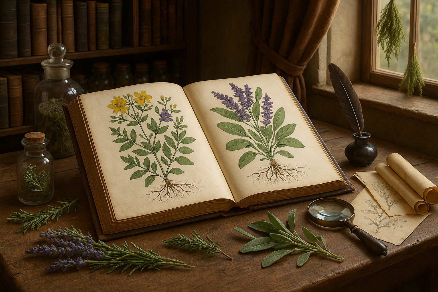
Toni Santos is a visual storyteller and archival illustrator whose work revives the elegance and precision of scientific illustrations from the past. Through a thoughtful and historically sensitive lens, Toni brings renewed life to the intricate drawings that once shaped our understanding of the natural world — from anatomical diagrams to botanical engravings and celestial charts.
Rooted in a deep respect for classical methods of observation and documentation, his creative journey explores the crossroads of art and science. Each line, texture, and composition Toni creates or curates serves not only as a tribute to knowledge, but also as a meditation on how beauty and truth once coexisted on the page.
With a background in handcrafted artistry and visual research, Toni merges historical accuracy with aesthetic reverence. His work draws inspiration from forgotten sketchbooks, museum archives, and the quiet genius of early illustrators whose hands translated curiosity into form. These visual relics — once found in dusty volumes and explorer journals — are reframed through Toni’s practice as enduring symbols of wonder and intellect.
As the creative force behind Vizovex, Toni curates collections, essays, and artistic studies that invite others to rediscover the visual languages of early science. His work is not just about images — it’s about the legacy of observation, and the stories hidden in ink, parchment, and pigment.
His work is a tribute to:
The discipline and artistry of early scientific illustrators
The forgotten aesthetics of exploration and discovery
The quiet beauty of documenting the natural world by hand
Whether you’re a lover of antique diagrams, a natural history enthusiast, or someone drawn to the timeless union of science and art, Toni welcomes you into a world where knowledge was drawn, not digitized — one plate, one specimen, one masterpiece at a time.